What's the big deal about calcium?

Almost all of the calcium in the body (~99%) is found in the bones (and teeth), where it is essential for building and maintaining bone to give the bone its strength. The remaining ~1% is dissolved in the bloodstream and other fluids where it is used for maintaining the function of the heart, muscles, blood and nerves. We continuously lose calcium each day through our skin, nails, sweat and urine.

What happens if your diet is low in calcium?

Our bodies cannot make calcium, therefore all our calcium requirements are provided by our diet. If our diets do not provide sufficient calcium, there will not be enough calcium available in the bloodstream for our bodies to function properly. This means some of the calcium crystals stored in the bone will dissolve to 'stock up' the calcium needed in the bloodstream. If your calcium intake remains too low, the calcium in your bone will continuously need to dissolve and be released into your bloodstream, and you risk losing bone strength.

Calcium absorption

As well as increasing your intake of calcium, it is also essential that the calcium is able to be absorbed by the body. Calcium absorption can be reduced by excessive alcohol or caffeine consumption, as well as consuming a diet high in animal proteins or drinking soft drinks 
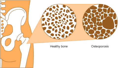
that contain phosphates. As we age, calcium is absorbed less effectively from the intestine thus we need to increase our intake of calcium to avoid losing bone density which may result in osteoporosis.

The greatest rate of bone growth is reached by puberty, and by age 30 we reach our peak bone mass (maximum bone density). The higher our peak bone mass, the better our bone health will be in the future. This is particularly important for women as rapid bone loss occurs during the menopause. As puberty is an essential time for determining your overall bone health, it is critical that children and teenagers get enough calcium.

Your daily calcium requirements depend on your age and sex. Less than half of Australian adults get their daily recommended intake of calcium.

Sources of calcium

Dairy
Dairy foods such as milk, yoghurt and most cheeses are calcium-rich and serve as the primary source of calcium in our diet. Calcium is also more easily absorbed from dairy products compared to other food groups.
Aim to eat 2-3 serves a day such as a glass of milk, a slice of hard cheese or a yoghurt.

Photo by adam morse on Unsplash
Canned fish
It's not only humans that have high levels of calcium in their bones. Consuming canned fish including the bones will also help you increase your calcium intake.
Try canned salmon or sardines!

Eat more fruit and veg!
Small amounts of calcium are also found in fruit and vegetables, particularly greens such as broccoli and kale. Nuts are also a great way to introduce more sources of calcium into your diet, particularly almonds.

Useful resources

Australian dietary guidelines 2013

Nutrient reference values for New Zealand and Australia

Arthritis WA

Calcium content of various foods

NHMRC Food for Health


Osteoporosis, osteoarthritis and rheumatoid arthritis – what's the difference?


When it comes to conditions affecting your bones and joints, many people confuse osteoporosis and certain types of arthritis including osteoarthritis. Although there are some similarities between these conditions, there are also many important differences.

Osteoporosis means 'porous bones' and is a condition in which the bones become weak and more prone to fracture due to a loss of bone density. Almost 1 in 10 Australians over the age of 50 years has osteoporosis (or osteopenia where bone density is low but not low enough to be classified as osteoporosis), with the condition being more prevalent in women than men. Osteoporosis can result is chronic pain, loss of independence and prolonged or permanent disability.

Arthritis
Arthritis is the term used for a group of conditions that affect the joints and surrounding tissues, causing pain, inflammation and damage to the joints. Arthritis is the major cause of chronic pain and disability in Australia with an estimated 3.85 million Australians affected. There are many different types of arthritis, the main two being osteoarthritis and rheumatoid arthritis.

Similarities between osteoporosis and arthritis
Much of the confusion between these two conditions is due to the similarity of their names – osteoporosis and osteoarthritis (by far the most common type of arthritis). It is true that osteoporosis and arthritis do share many coping strategies for the management of the disease. For example, many people with arthritis or osteoporosis benefit from exercise programs that emphasise a range of motion, stretching, strengthening and posture such as swimming, tai-chi and low-stress yoga. Physical therapy and rehabilitation may also be of benefit to some people. However, it is important that any exercise program is discussed with your doctor first to ensure your safety as your condition will affect your ability and you may need to compensate for certain movements.
Pain management strategies are also often similar where required, as is often the case for people suffering from arthritis, although less so for people with osteoporosis other than when in recovery from a fracture.

Other than these similarities in coping with and managing your disease, osteoporosis and arthritis are very different conditions.

Differences between osteoporosis, osteoarthritis and rheumatoid arthritis
Despite the similarity in name, osteoporosis and osteoarthritis are completely different conditions in terms of development, symptoms, diagnosis and treatment.

Even within arthritis, both osteoarthritis and rheumatoid arthritis are very different diseases. They may share some similar characteristics, but each has different symptoms and requires different treatment, thus an accurate diagnosis is essential. The primary difference between osteoarthritis and rheumatoid arthritis is the cause of the joint symptoms. Joint damage in osteoarthritis is a result of mechanical wear and tear that breaks down the cartilage between the joints, causing the bone ends to rub together. Rheumatoid arthritis on the other hand is an autoimmune disease where the body's own immune system attacks the tissue lining the joints. Both result in inflammation, pain and swelling of the joint.

Characteristic
Osteoporosis
Osteoarthritis
Rheumatoid arthritis
Characteristic of disease
Loss of bone density causing weak and brittle bones
Chronic joint disorder of the cartilage
Autoimmune disease that attacks the tissue lining the joint
Age of onset
Usually later in life
At any time in life
Speed on onset
Over years
Over years
Weeks to months
Who is affected?
1 in 10 Australians over 50 years of age
1 in 11 Australians
2 in 100 Australians
Affect on joints
N/A
Pain and stiffness, usually located in one set of joints on one side of the body, although symptoms may spread
Pain, swelling and stiffness affecting joints symmetrically (on both sides of the body)
Affect on bones
Loss of bone density causes bones to become weak and brittle and prone to fractures.
Bone ends rubbing against one another may result in bone projections known as bone spurs (osteophytes)
N/A
Systemic symptoms
N/A
N/A
Fatigue and general feeling of being unwell
Long-term impact
Increased risk to fractures and fracture cascade. Chronic pain, inability to walk, change in posture, disability, loss of independence.
Difficulty in performing everyday activities
Bone erosion, irreversible joint damage and permanent disability.
Diagnosis
May progress undetected for years until a fracture occurs. Diagnosed using a bone mineral density scan.
Physical examination, x-rays, blood tests (to rule out other types of arthritis)
Physical examination, blood tests, x-rays
Treatment
Lifestyle changes including fall and fracture avoidance, increase in calcium and vitamin D levels, regular exercise. Medications to promote bone formation and prevent bone degradation.
Analgesics, NSAIDs, Exercise and physical therapy, joint replacement therapy
Exercise and physical therapy, NSAIDs, Corticosteroids, DMARDs


Useful Resources

AIHW
http://www.aihw.gov.au/arthritis-and-musculoskeletal-conditions/

Osteoporosis Australia

Artritis WA
https://www.arthritiswa.org.au/

What is Rheumatoid arthritis?

What is rheumatoid arthritis?
Rheumatoid arthritis (RA) is an chronic autoimmune disease which causes marked inflammation of the joints. The body's own immune system attacks the synovial membrane (the tissue lining of the joint), leading to inflammation and pain as well as swelling and stiffness (See Figure 1).

Figure 1: Diagram showing difference between a healthy joint and RA (http://www.aihw.gov.au/rheumatoid-arthritis/about/)


The synovial membrane is usually very thin, and produces the fluid to lubricate and nourish the joint tissue. However, in RA the synovial membrane becomes thick and inflamed, resulting in unwanted tissue growth which may lead to bone erosion, irreversible joint damage and permanent disability. RA usually affects the smaller joints, most often the joints in the hands, and also the feet, but can also affect the larger joints in the knees and hips. As a systemic disease, it affects the whole body and can also lead to problems with the heart, respiratory system, nervous system and eyes.

What causes RA?
The precise cause of RA is not well understood, although it is known that there is a strong genetic component, contributing to 50-60% of the risk for developing RA. Smokers and people who have a family history of RA also have a higher risk of developing the disease.

Who gets RA?
According to the Australian Institute of Health and Welfare, approximately 407,900 Australians (approximately 2% of the total population) have RA, based on self-reported data from the ABS 2014-15 National Health Survey. RA can affect anyone at any age although is most common in those over 65 years, and has a slightly higher prevalence in women (1.7%) compared to men (1.4%). Over an ~10 year period between 2005-06 and 2014-15, there was a 59% increase in the rate of hospitalisation due to RA, primarily due to female patients.

Symptoms and limitations caused by RA
The most common symptoms of RA include joint pain, swelling, stiffness and tenderness, and usually affect the same joint on both sides of the body at once. Systemic symptoms may include fatigue, fever, weight loss, anaemia, inflammation of the eyes or lungs, and subcutaneous nodules (bumps under the skin).
Quality of life can be severely affected for people with RA. Even in the early stages of disease, severe limitations may occur, which then worsen over time. The cause of the most severe limitation is most commonly reported to be joint damage in the wrist. There is also a high prevalence of anxiety, depression and low self-esteem amongst people with RA.
Another major consequence of this condition is work disability, with only 80% of patients being able to continue work 2 years after disease onset, and 68% at 5 years.

Diagnosis of RA
RA is diagnosed through a physical examination of your symptoms by your doctor and various tests including blood tests for inflammation and for rheumatoid factor, as well as x-rays to look for joint damage. During the early stages of disease these tests may come up negative, and your symptoms may be similar to other types of arthritic disease. Hence it can be difficult and time-consuming to diagnose RA, and if your doctor suspects you are affected you should be referred to a specialist known as a rheumatologist.

Treatment for RA
Currently there is no cure for RA, however there has been dramatic improvements in treatment of RA over recent years which are particularly helpful for people in the early stages of the disease. Treatment depends on your symptoms and their severity, and your rheumatologist may need to trial several different treatments in order to determine which one will work best for you. Treatment options may include:
  • Regular exercise
  • Non-steroidal anti-inflammatory drugs (NSAIDs)
  • Corticosteroid medications
  • Disease-modifying anti-rheumatic drugs (DMARDs)
  • Biological DMARDs such as tumour necrosis factor medications
Useful resources

AIHW

Arthritis WA

Osteoporosis - the silent disease

What is osteoporosis?
In osteoporosis (which means 'porous bones') bones become weak and fragile which means that even a minor bump or accident can result in a broken bone known as a minimal trauma fracture. These can result in chronic pain, disability, loss of independence and premature death. Brittle bones usually result from a decrease in bone mineral density (BMD) and changes in bone quality (See Figure 1). Decreased bone density occurs when bones lose minerals including calcium faster than the body is able to replace them.

Figure 1: Difference between healthy bone and osteoporosis. Image sourced from AIHW (http://www.aihw.gov.au/osteoporosis/what-is/)


Osteopenia is a related condition where the mineral density is lower than normal but not enough to be classified as osteoporosis.

The 'fracture cascade'
The risk of future fractures increases with each new fracture, known as the 'cascade effect', with women who have a spinal fracture 4x more likely to have another fracture within a year. This number increases the more fractures an individual experiences, with an 11x greater risk of fracture in people who have already experienced 3 or more compared to someone who hasn't had one.

Who is at risk?
Older people and post-menopausal women have a greater risk of developing osteoporosis or osteopenia. It is a common disease in Australia with almost 1 in 10 Australians over the age of 50 have either osteoporosis or osteopenia, with women over the age of 50 years 4x as likely to be affected by these conditions than men of the same age. This is because of the rapid decline in oestrogen levels during menopause which results in an increased rate of loss of bone calcium and other minerals. This results in approximately 2% bone loss per year occurring for several years post menopause. The rate of hospitalisation for minimal trauma fracture is 2.6x higher for women over 50 compared to men.

Risk factors for osteoporosis
There are a number of risk factors associated with the development of osteoporosis including:
  • Increase in age
  • Being female
  • Having an early menopause or being postmenopausal due to the rapid decline in oestrogen levels
  • Family history of osteoporosis
  • Reduced levels of oestrogen
  • Low calcium intake
  • Low levels of vitamin D (which your body needs to absorb calcium)
  • Low body weight or small frame
  • Smoking
  • Lack of physical activity
  • Excessive alcohol consumption
  • Medical history including: malabsorption disorders (e.g. coeliac disease), some hormonal disorders (e.g. thyroxine excess) or long-term use of certain medications (e.g. corticosteroids)
Symptoms and resulting issues
Osteoporosis usually has no signs or symptoms until a fracture occurs, hence why it is known as a 'silent disease'. Osteoporosis (and the associated fractures) can result in:
  • Increased risk of fractures
  • Loss of height
  • Severe back pain
  • Change of posture
  • Muscle weakness
  • Spinal bone deformity
  • Impaired ability to walk
  • Chronic pain
  • Loss of independence
  • Premature death
Diagnosis of osteoporosis
Osteoporosis is often referred to as a silent disease as it can progress undetected for many years until a fracture occurs. It is diagnosed using a bone mineral density test (also known as a 'dual energy X-ray absorptiometry (DXA) scan') to measure the bone mineral density in the hips and spine. The results of the scan will be compared to the average BMD of healthy adults and expressed the as a T-score. The range of T-scores will determine whether the individual's bone density is normal or an indication of osteopenia or osteoporosis (See Table 1).

Table 1: How T scores relate to bone density health

Condition
T score
Normal
1 to -1
Osteopenia
-1 to -2.5
Osteoporosis
-2.5 or lower

Prevention and management of osteoporosis
The best prevention methods depend on your bone density and T-score results. If the test shows normal bone density recommendations for maintaining good bone health include exercise and adequate intake of calcium and vitamin D. If the test shows osteopenia further lifestyle changes may be required as well as a follow up bone density scan in 1-2 years to monitor your bone health. If the results show osteoporosis it means you are at risk of fracture and important lifestyle changes and fall prevention measures need to be taken. You'll also likely start treatment to prevent further bone loss and fractures and require a follow up test in a year's time. It is essential that any osteoporotic fractures are identified and treated as quickly as possible in order to stop the fracture cascade.

Lifestyle changes
Additional modifications to day to day behaviour can prevent or slow down the development of osteoporosis, or lessen the effects of the disease. These include:
  • Increasing calcium intake through diet or supplements
  • Increase vitamin D levels with sunlight exposure or supplements
  • Partaking in regular weight-bearing exercise
  • Stopping smoking
  • Decreasing alcohol intake
  • Possible changes in your normal medications
  • Adjusting behaviour to avoid fractures wherever possible
Medications
Bone tissue is constantly broken down (by osteoclasts) and renewed (by osteoblasts) in a carefully balanced cycle. Osteoporosis unbalances this cycle. Medications for managing osteoporosis inactivates osteoclasts to stop bone tissue break down, but allows osteoblasts to continue forming new bone tissue. This results in a gradual increase in bone tissue density over time.

Useful resources

AIHW

Osteoporosis Australia

Artritis WA

Osteoporosis Medications

What is osteoarthritis?

What is osteoarthritis?
Osteoarthritis is a degenerative condition that usually gets worse over time. As the cartilage that covers the ends of the bones in the joints degrades (breaks down), the bones start to rub together causing pain and sometimes swelling (See Figure 1). As joints become stiff there is a loss of motion that combined with the pain makes performing everyday activities difficult.

Figure 1: Difference between a normal bone joint and osteoarthritis. Image sourced from AIHW (http://www.aihw.gov.au/osteoarthritis/what-is-osteoarthritis/)


Osteoarthritis mostly affects the the hands, spine, hips, knees and ankles, and usually develops in joints that are injured due to repeated overuse. It is the predominant condition leading to hip and knee replacement surgery in Australia, with an 29% rise in the rate of total knee replacements and a 31% rise in hip replacements occurring over the last ~10 years.

Symptoms
Although the symptoms of osteoarthritis can vary between people, and over time for the affect person, the main symptoms of include:
  • Joint pain (particularly pain that worsens following exercise or immobility)
  • Joint stiffness
  • Swelling
  • Limited joint movement
Who gets Osteoarthritis?
An estimated 1 in 11 Australians (approximately 2.1 million people in 2014-2015) are believed to have osteoarthritis, with approximately two thirds of these being female.

Risk factors
There are a number of risk factors that contribute to the onset and progression of osteoarthritis including:
  • Being female
  • Joint injury or trauma (i.e. dislocation or fracture)
  • Joint misalignment
  • Repetitive joint-loading tasks (such as kneeling or squatting)
  • Being overweight
  • Family history of osteoarthritis
Diagnosis
Osteoarthritis is usually diagnosed through a physical examination of your symptoms and the affected joints. X-rays may be used to observe any narrowing or changes to the shape of the joint but cannot determine the amount of pain or problems you experience in this joint. Blood tests may be used to rule out other types of arthritis.

Treatment for osteoarthritis
Treatment options depend on which joints are affected and to what severity. It make take some trials before you find the best treatment for you as an individual. Example treatments include:
  • Analgesics (pain relief such as paracetamol)
  • Topical creams
  • Nonsteroidal anti-inflammatory drugs (NSAIDs)
  • Exercise and physical therapy
  • Joint splinting
  • Joint replacement surgery if all other therapies are no longer helping
Useful resources

AIHW

Arthritis Australia

Ovarian cancer in Australia

Incidence rates of ovarian cancer

Ovarian cancer is the eighth most common cancer in Australian females and the second most commonly diagnosed gynaecological cancer after uterine cancer.

Approximately 1,580 Australian women are expected to be diagnosed with ovarian cancer in 2017, accounting for 2.5% of all new female cancer cases. The estimated age-standardised incidence rate for 2017 is 10.8 cases per 100,000 females, up from 10.6 cases per 100,000 females in 2013 (Figure 1). The majority of these women diagnosed are likely to be diagnosed with an advanced stage of disease as early-stage ovarian cancer often does not present with symptoms.

The risk of ovarian cancer increases with age up until the age group 70 – 95+ where the incidence begins to decrease. The average age at diagnosis is 63 years, with a 1 in 122 risk of developing ovarian cancer before the age of 75, and a 1 in 81 risk before the age of 85.

Figure 1: Estimated incidence rates and mortality rates for ovarian cancer in 2017


Mortality rates for ovarian cancer

Ovarian cancer is the sixth most common cause of cancer death amongst Australian women, with an estimated 1,047 deaths (accounting for 5.1% of all female cancer deaths) expected in 2017. The age-standardised rate (ASR) for mortality was 6.8 deaths per 100,000 cases in 2014, a decrease from 9.3 deaths per 100,000 females in 1968 (Figure 1).

The 5 year survival rate between 2009-2013 was 44.4%, up from 34.1% between 1984-1988 (Figure 2). The prognosis (outlook for survival) depends on the type and stage of cancer at diagnosis as well as general health. Where ovarian cancer is diagnosed early, over 80% of women are likely to survive for over 5 years, however only approximately 30% of women diagnosed at advanced stages will survive for more than 5 years.

Figure 2: 5-year relative survival from ovarian cancer

Resources
Cancer in Australia 2017

Summary of AIHW Cancer in Australia 2017 report

Ovarian cancer

What is ovarian cancer?

Ovarian cancer occurs when cells in one or both of the ovaries begin to grow abnormally and develop into cancer. There are four main types of ovarian cancer that are named after the cell types where the cancer originates:

Epithelial ovarian cancer
  • Originates in the epithelium (outer cells of the ovary) and accounts for ~90% of cases.
Borderline or low malignant potential (LMPO) tumours
  • Less aggressive epithelial tumours with a generally favourable prognosis for the women affected.
Germ cell ovarian cancer
  • Arises in the cells that mature into eggs (ova) and accounts for ~5% of ovarian cancer cases, particularly affects women under 30 years.
Sex-chord stromal cell cancer
  • Begins in the ovarian cells that release female hormones, also accounts for ~5% of cases and can affect women of any age.
Cause of ovarian cancer

There are a number of factors which may increase a woman's risk of developing ovarian cancer, such as:
  • Family history
  • Being of Northern European or Ashkenazi Jewish descent
  • Alterations in BRCA1 or BRCA2 genes
  • Being over the age of 50
  • Late menopause
  • Early onset of menstruation (<12 years)
  • Infertility, never having children or having their first child after the age of 30 years
  • Never taking oral contraceptives
  • Having fertility treatment or oestrogen-only hormone replacement therapy
  • Smoking
  • Obesity
Symptoms of ovarian cancer

Although there are no obvious signs of ovarian cancer, you may experience some of the following symptoms:
  • Abdominal bloating
  • Difficulty eating/feeling full quickly
  • Frequent/urgent urination
  • Constipation
  • Indigestion
  • Back, abdominal or pelvic pain
  • Menstrual irregularities
  • Fatigue
  • Pain during sex
Diagnosis

There is currently no screening option for ovarian cancer in Australia. However, if you are experiencing symptoms associated with ovarian cancer it is advised that you discuss your concerns with your doctor who may suggest various tests to detect cysts, tumours or other abnormalities. These may include:

Physical examination
  • External abdomen examination and internal vaginal examination.
Blood tests
  • Your blood will be tested for tumour markers, in particular the protein CA125 which may be elevated in women with ovarian cancer.
Ultrasound
  • A transvaginal ultrasound (TVU) where an ultrasound probe to be inserted into your vagina to relay an image of the ovaries.
Other Imaging scans
  • Other tests may include CT scans, external ultrasounds, abdominal x-rays or an MRI
If the tests suggest that there are abnormalities present, a biopsy (tissue sample) may be taken to confirm cancer diagnosis.

Staging

There are often few signs of ovarian cancer in its early stages, usually only presenting once the cancer has spread. The stage of ovarian cancer is determined using the International Federation of Gynaecology and Obstetrics (FIGO) system. The FIGO system records whether the cancer remains contained within the ovaries or the extent of which it has spread to other pelvic structures or into the abdominal lining.

Treatment

Your treatment team generally consists of your GP as well as a number of specialists including a gynaecological oncologist and possibly a medical oncologist or radiation oncologist, as well as other healthcare professionals. The best treatment option depends on the type and stage of the ovarian cancer as well as general health.

Surgery
  • Surgery is usually the main treatment option for ovarian cancer, particularly when the cancer is still localised. The initial operation is known as a laparotomy, where a long vertical cut is made in the abdomen in order for the surgeon to locate and remove as much of the tumour as possible. At the start of the laparotomy a biopsy (known as a frozen section) is taken to confirm diagnosis. During the operation most women will have their ovaries, Fallopian tubes, uterus, omentum (fat pad around the abdominal organs), appendix and some lymph nodes removed, as well as part of the bowel in some instances. Biopsies from the removed tissues are examined to enable the gynaecological oncologist to learn more about the type and extent of the cancer and determine the best course of action for further treatment.
Chemotherapy
  • Most women with ovarian cancer also undergo chemotherapy to slow or cease the growth of remaining cancer cells. Chemotherapy is most effective when the cancer is small and the cells are actively growing and is commonly used soon after surgery to remove remaining cancer cells. It may also be used as a first line of treatment in cases of widespread disease, or in situations where ovarian cancer has returned. Chemotherapy can damage non-cancerous cells and cause side effects. Advice for reducing of managing these side effects can be found here.
Radiotherapy
  • Radiotherapy is less often used as a treatment for ovarian cancer but may be used in cases where the cancer is confined to the pelvic cavity, or in cases of advanced ovarian cancer to reduce the size and relieve symptoms.
Palliative care
  • Palliative care may also be used in an attempt to alleviate symptoms, improve quality of life and slow the spread of ovarian cancer
Useful resources
Cancer Council Australia

Ovarian Cancer Australia

Uterine cancer in Australia


Uterine cancer is the most commonly diagnosed gynaecological cancer in Australia and is expected to remain the 5th most commonly diagnosed of all cancers amongst females in Australia for 2017.

In 2017 it is estimated that 2,861 new cases of uterine cancer will be diagnosed in Australia, accounting for approximately 4.6% of all new female cancer diagnoses. Over the last 35 years the number of new cases of uterine cancer has increased from 942 in 1982 to the estimated 2,861 new cases expected for 2017.

The incidence rate for uterine cancer increases with age from age group 20-24 until age group 65-69 after which it begins to decrease again (Figure 1). Overall, the age-standardised incidence rate for uterine cancer is 19 cases per 100,000 females, an increase from 14 cases per 100,000 females in 1982 (Figure 2). The risk of a female being diagnosed with uterine cancer by the time she turns 85 years old is estimated to be 1 in 42 for 2017.

Figure 1: Estimated incidence rates and mortality rates for uterine cancer in 2017


It is estimated that uterine cancer will be the 13th leading cause of cancer death for females in Australia in 2017, down from the 12th leading cause in 2014. This corresponds with a decrease in the number or deaths resulting from uterine cancer from 494 in 2014 to an estimated 453 deaths in 2017, which will likely account for 2.2% of all female cancer deaths.

Likewise, the age-standardised mortality rate is estimated to decrease from 3.4 deaths per 100,000 females in 2014 to 2.8 deaths per 100,000 females (Figure 2). However, mortality rate for uterine cancer generally does increase with age, particularly for those over the age of 60 years old (Figure 1), with an estimated risk of 1 in 259 for a female dying from uterine cancer by her 85th birthday in 2017.


Figure 2: Age-standardised incidence and mortality rates for uterine cancer


From 2009-2013 females diagnosed with uterine cancer had a 5-year survival chance of 83.2%, an improvement on the 5-year relative survival of 75% between 1984-1988.

Figure 3: 5-year relative survival from uterine cancer


Resources

Cancer in Australia 2017

Summary of AIHW Cancer in Australia 2017 report


Popular Posts